|
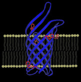
The transmembrane domain of a nonspecific β-barrel pore located in the outer membrane of bacteria, Outer Membrane Protein A (OmpA),
is displayed on the left (PDB 1QJP) along with a cartoon representation of the lipid bilayer.
The five native tryptophan residues found at the bilayer-water interface are shown in red.
OmpA serves as our model β-barrel integral protein for fundamental studies on membrane protein folding. Click here to read more... |

|
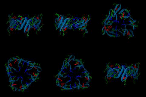
A postulated hexameric β-barrel structure of a human antimicrobial peptide in the defensins family,
HNP3, is shown on the left; this structure is composed of three dimer units (PDB 1DFN) and may be functionally relevant for membrane disruption.
In each monomer, the single tryptophan residue is shown in red, cationic residues are shown in green, and anionic residues are shown in blue.
The goal of our research is to probe molecular changes in antimicrobial peptide structure upon membrane association and subsequent membrane disruption. Click here to read more... |

|
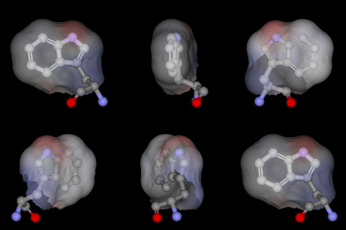
The highly buried, single tryptophan residue in the blue copper protein, azurin,
is highlighted on the left (PDB 4AZU). Azurin continues to be an important system for the study of electron transfer reactions;
as a result, we are utilizing this protein as a model system to study radical intermediates in long-range electron transfer reactions.
An important aim of this research project is to elucidate the structure and kinetics of biological radicals,
namely aromatic amino acid radicals, using an array of powerful spectroscopic tools.
Click here to read more... |

|
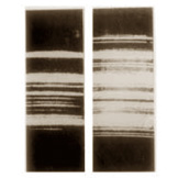
Chandrasekhara Venkata (C.V.) Raman received the Nobel Prize in Physics in 1930 “for his work on the scattering of
light and for the discovery of the effect named after him.” The first spectrum taken in 1928 by C.V. Raman and his
colleague K.S. Krishnan is shown on the left (from R. Singh, Phys. Perspect. 2002). The left photograph shows the
incident light from a mercury arc lamp after passing through a blue filter; the right photograph shows the same spectrum
after passing through liquid benzene. Click here to read more... |


